Introduction

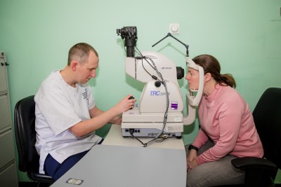
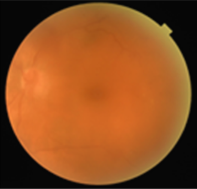
When you attend for your diabetic eye screening appointment, a retinal camera is used to take a detailed photograph of the back of your eyes (the retina). During your appointment, drops will be inserted into your eyes, this is to dilate your pupils (make them nice and big) and allows us to capture multiple detailed images. These images are then reviewed to check for any signs of diabetic retinopathy.
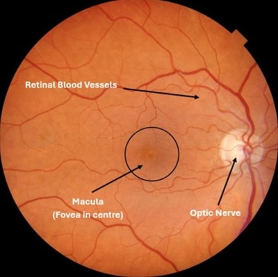
This is the light sensitive layer of tissue at the back of the eyeball that receives images and sends them as electric signals through the optic nerve to the brain.
This is part of the retina and is responsible for our central vision (what you can see directly in front of you) the majority of the colour vision and also the fine details of what we can see.
This is at the back of each eye and contains millions of nerve fibres that send visual messages to your brain, the brain then converts the signals into images that helps you to see.
These are the blood vessels inside the retina that can be observed live, these arteries and veins play a crucial role in supplying vital oxygen and nutrients to the retina.
Diabetic retinopathy can affect anyone who is living with diabetes and can occur by having uncontrolled high blood sugar levels (HBA1c) over a prolonged period, this can damage the blood vessels inside the retina. It causes the tiny blood vessels within the eye to become damaged, get blocked and bulge, leak fluid or bleed and if left untreated this can lead to sight loss or even blindness.
Diabetes can affect your eyes in many ways, and the severity is recorded in the main stages below, which you will see in your results letter.
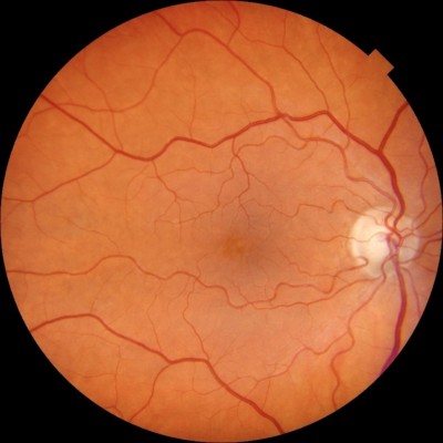
This means that your test results showed there were no signs of diabetic retinopathy detected in your eyes, please continue to effectively control your diabetes.
You will be asked to attend screening in either one or two years. If there are no signs of diabetic retinopathy on two consecutive visits, then you will be asked to attend eye screening every two years.
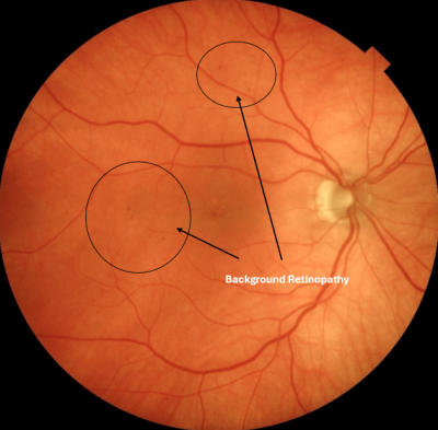
This is called Background Retinopathy and means your test results show diabetes has started to affect the blood vessels in the retina. At this stage you do not require any treatment, and it does not usually impact your vision, however it might progress and get worse if you do not control your diabetes. In the very early stages, some symptoms can resolve on their own, you can reduce the risk by effectively managing your diabetes.
You will be asked to attend screening in one year.
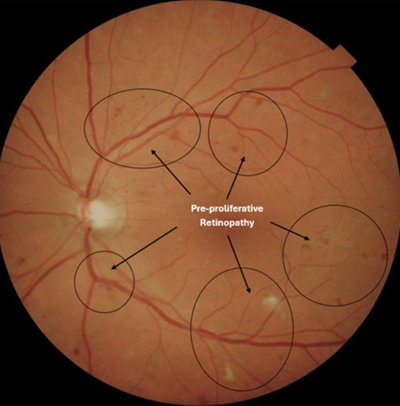
This is called Pre-proliferative Retinopathy and means your test results show more serious and widespread changes have been found to the blood vessels in the retina. This can increase the risk of vision loss, it is therefore very important that you take steps to manage your diabetes effectively, to try and reduce further changes from developing.
You will be asked to attend screening more frequently, usually every 3 or 6 months so we can closely monitor your eyes. Depending on the severity of the changes, you may be referred into the hospital eye services to help lower the risk of deterioration to your vision.
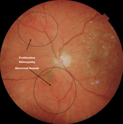
This is called Proliferative Retinopathy and means your test results show scar tissue and new blood vessels have developed on the retina, these vessels are weak and can bleed very easily, which means your vision could be seriously affected. We will refer you to the hospital eye services and you will be seen within three weeks from your screening appointment. It is very important to attend this appointment as you may need treatment to prevent further damage to your vision.
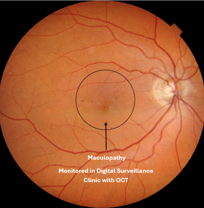
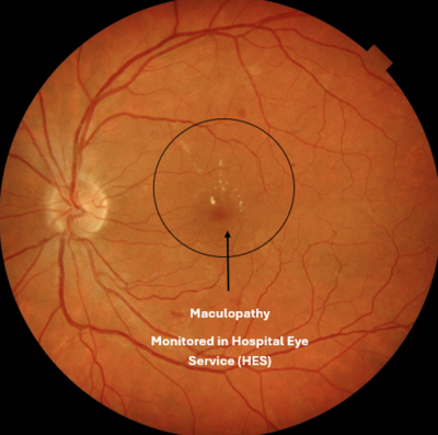
There is also an “M” in your eye screening results, this stands for Diabetic Maculopathy. The macula is the small central part of the retina which controls your sharp vision and helps you to see things clearly. If your results show M0 this means there are no signs of maculopathy, if your results show M1 this means maculopathy has been found.
Maculopathy is when diabetic changes occur in the macula area and consequently it could affect your vision. You would need to be screened more frequently or be referred to hospital eye services and offered treatment to help reduce the risk of vision loss.
People with maculopathy can often experience blurred or fuzzy central vision, difficulty seeing in dim/low light (especially at nighttime), visual distortion (straight lines can appear wavy), difficulty when trying to focus, light and glare sensitivity and colours may appear faded/less vibrant.
If your results show M1, this means you will be referred into our digital surveillance pathway and monitored more frequently using our Optical Coherence Tomography (OCT) equipment. This can be carried out in the retinal screening clinics at either Hunden’s Lane in Darlington, University Hospital of North Durham, Chester- Le-Street, Shotley Bridge, Sedgefield and Bishop Auckland Hospital or you may be referred to the Hospital Eye Service for closer investigation / treatment if required.
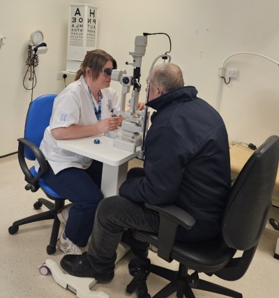
Sometimes it is very difficult to get good quality images, this can be for various reasons, for example if you have a dense cataract, unable to position at the camera or follow instructions. In these circumstances if we are unable to take good quality photographs of the back of your eyes, you may be referred to our slit lamp clinic. The slit lamp is a special microscope where a beam of light (a slit) is used to get a live, detailed view of the back of your eyes and allows us to examine and determine any abnormalities or changes that may have been caused due to diabetic retinopathy.
If we see changes that need further screening, you will be transferred to our Digital Surveillance pathway and be invited more frequently to assess and monitor these. This may be on a three or six month recall. Depending on the changes we may take more images to view a wider area of the retina.
As part of this appointment, we may also take on Optical Coherence Tomography (OCT) scan. This is a piece of technology that allows us to take a 3D image and view the deeper layers, beneath the surface of the eye. It’s a very quick, painless and non-invasive (does not touch your eye) procedure, that uses light wave technology, allowing us to have an incredibly accurate and detailed, cross-sectional view of each distinctive layer of the retina and optic nerve. It can help us to identify features of treatable diabetic retinopathy and maculopathy, as well as a range of other eye conditions, much more precisely than a 2D digital retinal image.
The OCT scan is very similar to having your photographs taken. We will insert drops into your eyes and you will be asked to have a seat in the waiting area whilst your pupils dilate. Once the drops have worked you will be invited back into the screening room and asked to sit in front of the OCT machine, resting your chin on the chinrest and forehead against the headrest bar. Once we have made sure you feel comfortable and you are in the correct position, you will be asked to look at a stationary light, and we will start the scan. You will see a red bar, move from top to bottom, scanning across the back of the eye, it only takes a few seconds for each eye. To achieve the best results from your scan, try to keep as still as possible, keep your eyes wide open, look at the stationary light and try not to blink.
Please note: not all our clinics have the OCT available, so therefore you might need to travel to have this appointment carried out.
The are no known complication or side effects from people who have had an OCT scan. It’s a very safe, non-invasive and painless procedure. The appointment will last approximately 30-40 minutes.
For any serious/widespread eye changes found, you will be referred to the hospital eye services for further assessment and possible treatment, depending on the severity this could include retinal laser treatment, Anti-VEGF injections or surgery.
The key to reduce the risk of developing diabetic retinopathy or preventing it from getting worse is being able to keep your HbA1c (blood sugar) levels under control and taking proactive steps to manage your diabetes and a healthy lifestyle.